Case Study
Patient:
An 18 year old male was found collapsed face down in the garden with multiple stab wounds to his head, neck thorax and back. On arrival by the paramedics and physician staffed retrieval service, there was evidence of massive blood loss. IV access was obtained and pressure applied to the bleeding sites. The patient had an initial GCS 15. 13 minutes after arrival of the pre-hospital team, the patient rapidly lost consciousness and developed a PEA arrest. A laryngeal mask was inserted and bilateral finger thoracostomies were performed. Volume resuscitation with PRBC and FFP was commenced. The decision was made by the retrieval physician to perform a clamshell thoracotomy. No obvious pericardial tamponade was found. There was no major pulmonary injury. Manual compression of the aorta was performed, and the patient regained spontaneous circulation with continue volume resuscitation. The patient was rapidly transferred to our Level 1 Trauma Centre.
Immediate Management:
The patient was taken directly to the operating room for exploratory thoracotomy and laparotomy. Injuries found included neck laceration and haematoma, multiple lacerations to the occiput, back, chest and abdomen, lung lacerations, diaphragm lacerations, liver lacerations, cephalic and axillary vein injuries. All visible bleeding was surgically controlled. The clamshell thoracotomy was closed and the abdomen temporarily closed with an Abthera VAC. The patient was then transferred emergently for angioembolization of muscular branches of the external carotid artery causing a massive haematoma in the neck. During this procedure, the patient suffered a cardiac arrest necessitating re-opening of the thoracotomy and evacuation of a tension haemothorax. The chest cavity was packed and temporarily closed utilising a VAC dressing. The patient was transferred to the ICU for ongoing resuscitation and rewarming.
In the first 24 hours, the patient required resuscitation with 78 units PRBC, 66 units FFP, 38 units apheresis cryoprecipitate, 3x pooled plates, 2000IU Prothrombinex.
Progress:
At relook laparotomy, there was evidence of colon infarction requiring a right hemicolectomy and formation of end ileostomy.
During the first week, the patient developed progressive multiorgan failure requiring respiratory and circulatory support as well as dialysis.
Wound Problem and Management:
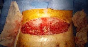
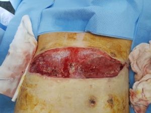
2 weeks after presentation, the thoracotomy wound developed purulent discharge and dehiscence. The patient was operatively debrided and copiously lavaged in the operating room every 3 days. The wound was managed with a VAC dressing system between debridements. Cultures from the wound debridements ultimately demonstrated multi-organism contamination including Mucor circinelloides, candida albicans, Enterobacter cloacae, Sternotrophomonas maltophila, and Pseudomonas aeruginosa. This massive wound contamination including traumatic mucormycosis was thought to be due to the significant multi-organ failure and immunosuppression. Figures 1 and 2 demonstrate the wound during initial debridements.
VAC Veraflo therapy was initiated after each subsequent debridement with the aim of reducing the contamination burden. In conjuction, broad spectrum antibiotics and anti-fungal agents were administered.
VAC Veraflo Therapy:
The Cleanse Choice foam was placed directly onto the wound after each operative debridement. Prontosan (polyhexanide) was utilised as the instillation fluid. “Fill assist” was used to determine the appropriate volume of instillation. The soak time was set as 15 min and the VAC as 2 hours.
Progress:
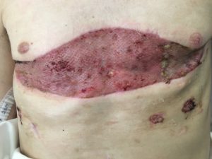
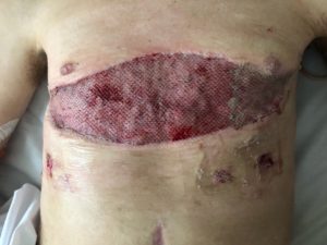
After a further 2 weeks of debridement and VAC Veraflo, wound cultures only demonstrated Pseudomonas aeruginosa. Instillation fluid was changed to Microdacyn. A further 4 weeks of VAC Veraflo with the Cleanse Choice foam was used to establish an infection-free, granulating bed (Figures 3 and 4). A split skin graft was performed to cover the wound. There was complete take of the skin graft (Figures 5 and 6)
Outcome:
The patient was discharged to rehabilitation after 3 months. He was markedly deconditioned, however all wounds were healed.
Conclusion:
This case demonstrates the utility of VAC Veraflo therapy to manage a complex, multi-contaminated wound in a critically ill trauma patient.

Appearance of wound after 4 weeks of VAC Veraflo therapy with Cleanse Choice

Appearance of wound after 4 weeks of VAC Veraflo therapy with Cleanse Choice
Appearance of wound week 2 after initial debridement
Appearance of wound week 1 after initial debridement
Appearance of wound 2 weeks after split skin grafting
Appearance of wound 1 week after split skin grafting