Case Study
Mr Mark Kim, Clinical Nurse Educator, St Vincent’s Hospital
Introduction:
This case study highlights the effectiveness of negative pressure wound therapy with instillation in preparing a wound bed for skin grafting in a patient with poor wound healing ability.
Patient History:
51 yo male with chronic bilateral lower limb cellulitis and lymphoedema with poor compliance. He had a motorbike accident more than 5 years prior which resulted in an upper thigh wound that was skin grafted. The grafted tissue was vulnerable to injury and a wound developed which became infected. When the patient was admitted into hospital on the 24th August, he presented with a deep thigh wound and multiple ulcers on his lower limbs. The patient was septic with high fevers and was initiated on IV antibiotics. Patient had hypoalbuminaemia and a very poor nutritional status which affected his ability to heal.
The lower leg ulcers were treated with a non-adherent content layer and Zetuvit to control wound exudate and the upper thigh wound was managed conservatively with standard wound care dressings.
Wound Treatment:
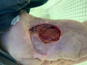
The upper thigh wound was covered with thick slough and on assessment the surgical team booked him in for surgical debridement. On the 8th September, the patient was taken into theatre for debridement and application of negative pressure wound therapy. Post debridement, the wound measured 15 x 10cm with a depth of 5cm. Swabs were taken which confirmed that the wound was colonized with Pseudomonas.
On the 11th September (Figure 1) the surgical team changed the treatment regime to negative pressure wound therapy with instillation and dwell (NPWTi-d), with a novel reticulated open cell foam dressing with through holes (ROCF-CC). The negative pressure device was set to instill normal saline with a 10 minute soak time every 4 hours, with the aim of keeping the wound bed clean and enhancing granulation tissue.
The instillation solution was changed to Microdacyn on the 17th September as there was concern of recurrence of Pseudomonas due to a mild odour from the wound at dressing change.
On the 27th September (Figure 3), the depth of the wound bed was reduced significantly and measured 1cm with healthy granulation tissue. The patient was continued with NPWTi-d with ROCF-CC, with only the solid cover layer used over the wound bed.
On the 7th October (Figure 4) the wound was reviewed by the surgical team and deemed to be ready for a skin graft. The patient was subsequently skin grafted 2 days later with negative pressure applied to the graft to ensure effective graft take.
Conclusion:
Considering the patient’s complex medical history, the utilization of NPWTi-d with ROCF-CC assisted to clean and granulate the wound for an effective graft take.
Figure 1: Prior to application of NPWTi-d with ROCF-CC

Figure 2: 23rd September

Figure 3: 27th September

Figure 4: 7th October
