Case Study
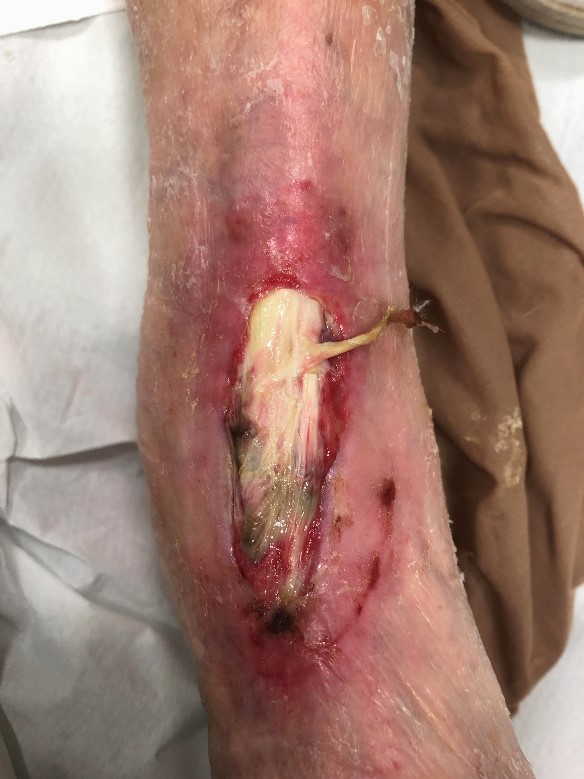
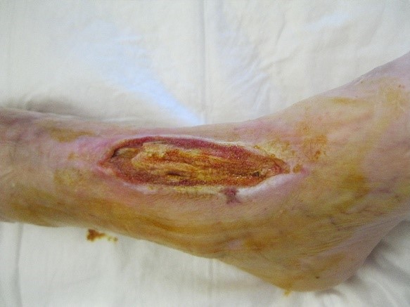
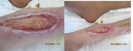
On Nov 26 2018 a 95 year old female is admitted due to a wound on the dosal side of her left foot, caused by trauma due to wheelchair (image 1). The GP already prescribed antibiotics several times, but the wound kept getting larger. The patient lives at a nursing home since 8 years, and due to her deteriorating wound, she was less mobile and more bound to her wheelchair. She has diabetes type 2, height 1,54m, weights 39,1kg and has a medical history of oesophagitis, hyperthyroid, osteoporosis and multi-infarct brain damage with repeating drop attacks, early stage dementia, for which her medication is adjusted.
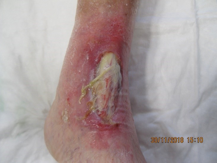
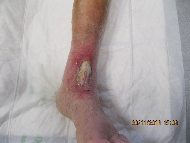
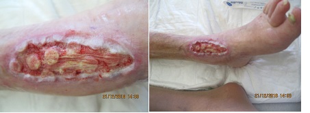
At admittance (image 2) we see a wound on the dorsal side of the left foot , exposed tendon with fibrin, inflammatory aspects around the wound, few exudate and a lot of pain during dressing changes. Wound cultures show staphylococcus aureus, CRP of 24,9 mg/dl and leucocytosis of 19,1. A CT angiograph of the lower limb vessels show a high grade stenosis of the left arteria poplitea.
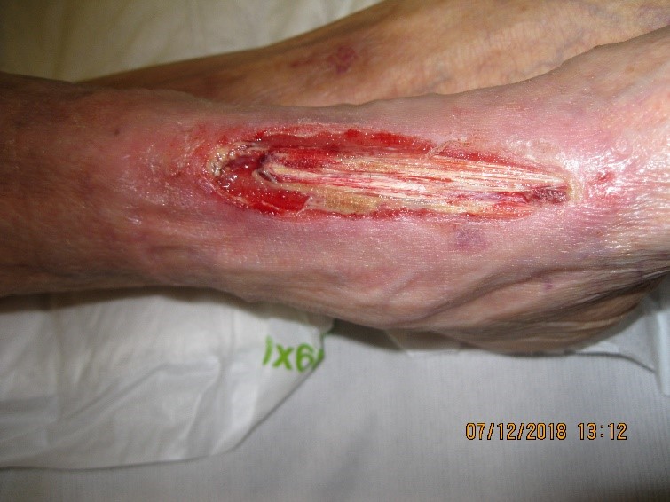
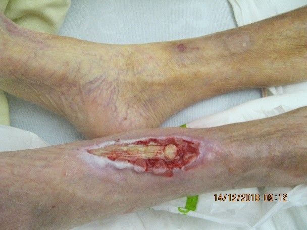
On Dec 6, 2018 a percutaneous transluminal angioplasty of the arteria poplitea sinistra is performed. Also a debridement of the ulcer is performed. (Image 3)
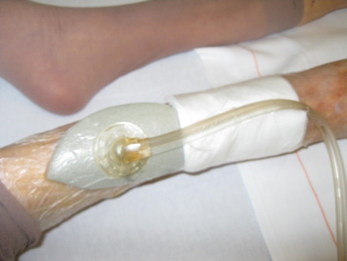
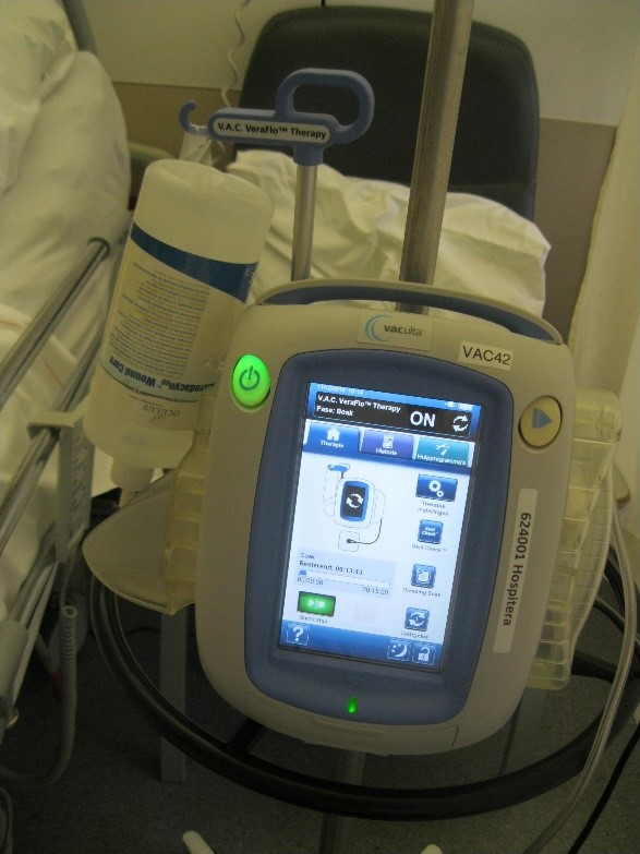
On Dec 9, 2018 VAC VERAFLO™ Therapy is initiated using the V.A.C.VERAFLO Cleanse Choice dressing. Instillation solution is Microdacyn with a soak time of 15 minutes, negative pressure at -125mmHg every 2 hours . Dressings changes two times per week. She also received systemic antibiotic treatment and nutrition supplements to gain strength. She was checked regularly for the glucose levels.
At the dressing changes (images 5, 6, 7) we noticed a progressive growth of granulation tissue, epithelialisation form the wound edges, few exudate and decreasing infection parameters. She is also stable for her diabetes. But we especially see an improvement in pain.
On Dec 24 the nurses form the nursing home are trained to perform VAC dressing changes in order to continue the therapy at the nursing home. She was discharged with an INFOVAC, intermittent therapy (5 min. on- 2 min. off) at negative pressure of -125 mm Hg . Dressing changes 2 times a week. Antibiotics was stopped. She continued with additional nutrition .
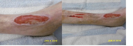
On Jan 8, 2019 (image 8) this photo was taken at the nursing home. The tendon in the wound is almost completely covered with granulation tissue. Unfortunately she has some blisters form the drape but this luckily healed quickly. There was frequent follow up including dressing changes, and the wound kept progressing (images 9 en 10).
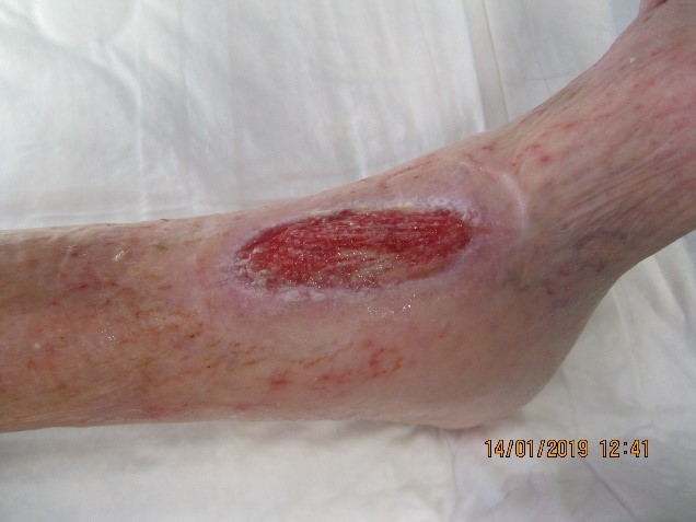
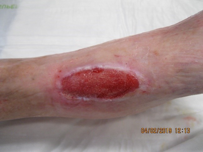
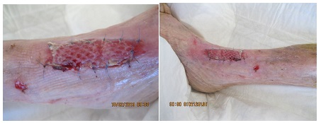
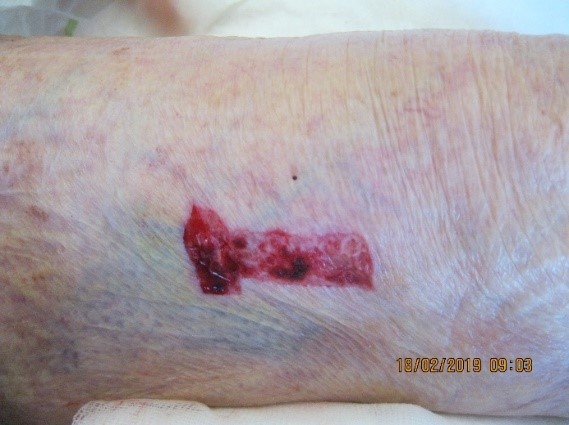
On Feb 14, 2019 (image 11 ) the wound is ready for a split thickness skin graft. There is good clinical and biochemical progress.
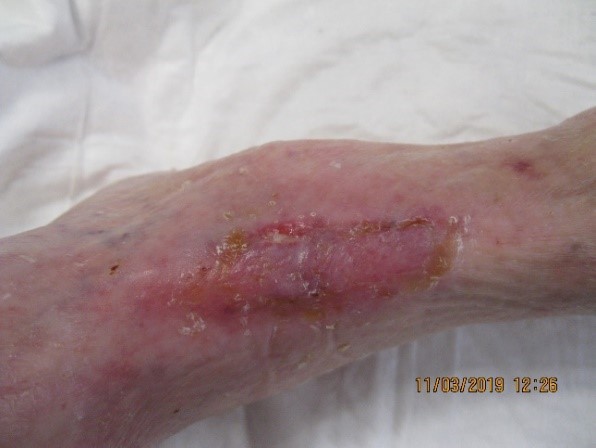
On March 13 the wound is fully healed and after several knee focused exercises, she is able to walk again using a rollator. She, her family and her GP did not expect this and were very happy with the result and the huge improvement in het quality of life.
