Case Study
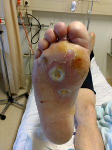
A 54-year-old man with no relevant medical history gets a blister during a soccer match on the right sole of his foot. Due to redness the GP prescribes antibiotic treatment, after which the wound is progressing. A week later new wounds appear on the foot and a local treatment and 2nd antibiotic treatment is started. Due to a serious deterioration of the wound (figure 1) the man presents himself in the emergency room.
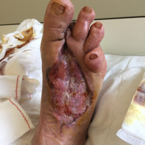
On admittance we notice a lot of inflammation at the foot. On the plantar side there are 2 ulcers of about 1 cm deep, with no apparent bone contact. On the dorsal side of the foot there is a lot of necrotic tissue. There is fluctuation on the dorsum of the foot. Blood results of the patient confirm the inflammatory status: leucocytosis of 12,19 and a CRP of 228mg/dl. Wound cultures show increasing numbers of staphylococcus aureus. There is a good capillary refill at the foot with palpable distal pulsations.
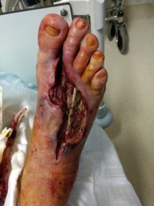
Immediate action is taken (December 2nd). An extensive surgical debridement and drainage of the pus and resection of the necrotic tissue is performed. The wound bed is irrigated thoroughly. Intravenous antibiotics is adjusted. In the following days more debridements are performed at the OR (Dec 4 and 6, figure 2) until all necrotic tissue is removed. At admittance, patient was diagnosed with Diabetes and insulin therapy is started and patient has been informed about the disease.
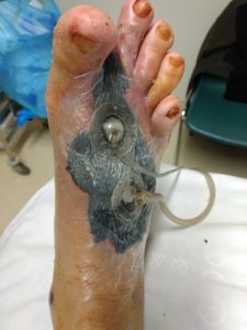
After the last debridement VAC VERAFLO™ Therapy is initiated (figure 3). Instillation solution is polyhexanide betaine (Prontosan) 20 ml and a soak time of 15 minutes, followed by VAC Therapy cycle, continuous therapy at -125mmHg. Wound edges are protected with Cavilon Advanced Skin Protectant. Dressing changed two times a week (Dec 11, 14 and 18, figure 4)
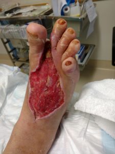
On December 20th, 13 days after starting VAC VERAFLO™ Therapy, a final debridement is performed of little necrotic tissue and resection of digit II. The wound is closed at the plantar side using a split thickness skin graft (figure 5). The clinical and biochemical results are positive.
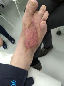
After 5 weeks the wounds have sufficiently healed and the patient is discharged on Jan 7, 2019. He has strict instructions not to stand on his foot and receives daily wound treatment by homecare nurses. Follow up on a weekly basis first and then bi- weekly in the multidisciplinary meeting on DFU. He receives adjusted orthopaedic shoes. After 4,5 months (figure 6) he can get back to work providing he wears the orthopaedic shoes.
In a situation of acute diabetic foot pathology, quick action is necessary. In the subacute phase, the use of VAC VERAFLO™ Therapy, is an important treatment tool and has a lot of value in healing complex diabetic foot wounds.
V.A.C. VERAFLO™ Therapy is a crucial part of the wound healing pathway in the multidisciplinary diabetic foot clinic of UZ Leuven.