Case Study
Necrotising fasciitis is a rapidly progressing often polymicrobial infection of the subcutaneous tissue and underlying fascia and is often triggered by, and rapidly spreads from, small lacerations on the affected area (Balci et al. 2018; Fink & DeLuca 2002). It is reported to be able to spread 1 inch per hour (Fink & DeLuca 2002). Management and prognosis is entirely dependent on the rapidity and adequacy of surgical debridement coupled with broad spectrum antimicrobial cover (Balci et al. 2018; Steinstraesser, Sand & Steinau 2009). Subsequently, after debridement, large wounds are inevitable and it has been shown in the literature that prior to or post grafting, negative pressure wound therapy (NPWT) facilitate granulation tissue formation and thus, wound healing (Balci et al. 2018; Braakenburg et al. 2006; Schiltz et al. 2019). Unfortunately, there are situations in practice where this is insufficient for wound healing such as in cases with poor vasculature or fixed wound contaminants.
As such, V.A.C VERAFLOTM Therapy is a novel device which optomises wound healing by combining the benefits of both NPWT and controlled instillation of topical solutions to wound beds. NPWT assists with reducing oedema and promoting tissue perfusion which allows the optimal substrate for the formation of granulation tissue. Additionally, delivery of topical solution, with or without soak time, assists with loosening contaminants, once again, promoting wound granulation. It is currently indicated in the use of chronic or acute wounds, wound dehiscence, partial thickness burns, ulcers, flaps and grafts.
In this report, we present a case of a young female presenting with necrotising fasciitis who benefited at multiple stages from the versatility of V.A.C VERAFLOTM Therapy.
Patient and diagnosis
A 44 year old female presented to a regional hospital emergency department with a two day history of general malaise, fever and labial abscess with purulent discharge and skin discolouration over her abdomen and pannus. This is on a background of being morbidly obese and suffering from type 2 diabetes mellitus on oral hypoglycaemics, congestive cardiac failure with an ejection fraction at worst of 21-30% (45-50% on repeat echocardiogram), severe biatrial dilatation and pulmonary hypertension, severe untreated obstructive sleep apnoea, polycythaemic and a history of previous unprovoked bilateral pulmonary emboli not on anticoagulation. Relevant medications included her heart failure medications carvedilol, spironolactone and dual antiplatelets. On presentation, the patient was haemodynamically unstable with the above findings and a diagnosis was promptly made of necrotising fasciitis involving the perineum, inguinal ligaments and lower abdomen.
Methods: Management and application of V.A.C VERAFLOTM Therapy
She was fluid resuscitated, given hydrocortisone and intravenous meropenum, vancoymcin, and lincomycin and rushed to theatre for emergency debridement. She was debrided twice on the first 24 hours of presentation, with the second planned return to theatre in order re-assess if further debridement was required. Her surgery required extensive debridement of abdominal, inguinal and perineal areas. The labial abscess with necrotic cavity was found to extend into the inguinal region reaching the abdomen. Severe tissue necrosis and abscess formation within the abdominal wall was debrided to normal fat and tissue – the rectus sheath remained intact. Post operatively, she was transferred to the intensive care unit where she remained on quadruple inotropes and vasopressors (adrenaline, noradrenaline, vasopressin and dobutamine). She was continued on her antimicrobials and commenced on continuous renal replacement therapy secondary to sepsis related acute renal injury.
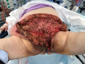
On day three of presentation, the decision was made to transfer her from the regional hospital to a tertiary centre for further management. On arrival, she was systemically well, off inotropes and vasopressors, however remained sedated, intubated and ventilated. She returned to theatre that day for review of wounds (Figure 1), requiring only minimal further debridement. The wounds were dressed with acticoat, black foam and V.A.C VERAFLOTM Therapy, 2 ports in synchronised setting with 65mL of solution soaking every 2 hours with pressure of 125mmHg. All antibiotics were subsequently ceased on day five post presentation as the patient was clinically stable and had already received sufficient coverage to treat the, now identified, coagulase negative staphylococcus grown on tissue and blood cultures. She was subsequently extubated and discharged to the ward on day six.

Figure 1. Abdomen and perineum after relook debridement, day three after presentation and prior to V.A.C VERAFLOTM Therapy.
The V.A.C VERAFLOTM Therapy dressing was changed a further two times on similar settings, and an additional NPWT dressing was added on the lower surface of the wound. A healthy granulating cavity was identified on day 9 since application of V.A.C VERAFLOTM Therapy. To assist with patient rehabilitation, a customised abdominal binder placed by occupational therapy and patient was mobilised with the assistance of physiotherapy.

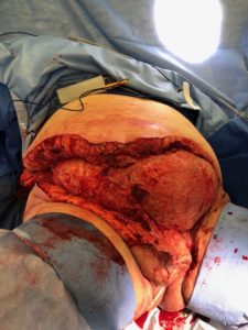
Figure 2. Wound after 17 days of V.A.C VERAFLOTM Therapy demonstrating healthy granulation tissue.
Results: Progress, discharge and follow-up
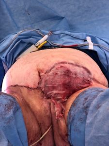
After 17 days of V.A.C VERAFLOTM Therapy, the wound was ready for application of split skin graft (Figure 2). Due to the weight of her pannus on the right side of her abdomen, the wound had started to dehisce. As such, the pannus was resected and that skin used as split skin graft to cover the rest of her wound (Figure 3). Cavities were packed with bactigras and V.A.C VERAFLOTM Therapy was placed over the graft for a further 10 days (Figure 4). Staples were removed 2 week post grafting. The patient was subsequently discharged for outpatient follow-up and daily simple wound dressings after 5 weeks of hospitalisation essentially independent with activities of daily living and graft well taken (Figure 5).

Figure 3. Abdominoplasty performed due to tension resulting in wound dehiscence of right sided abdominal wound. Split skin graft was placed over the remaining wound.

Figure 4. V.A.C VERAFLOTM Therapy in place over wound dehiscence and remaining grafted wound.

Figure 5. Split skin graft on left sided abdomen was observed to be taking well after 10 days of V.A.C VERAFLOTM Therapy and subsequent simple dressings.
Discussion
Rapid diagnosis and surgical debridement are the crucial elements in the successful immediate management of necrotising fasciitis. However, life saving debridement is only the beginning of patient care with the management of the resultant large skin defect, be it coverage or management of subsequent complications, needing to be considered.
Current literature suggests that NPWT can reduce the number of dressing changes, is more comfortable for the patient and can improve healing time by stimulating granulation tissue formation (Braakenburg et al. 2006; Maurya & Bhandari 2016; Morykwas et al. 1997). In more complicated wounds, NPWT with dwell time similar to the V.A.C VERAFLOTM Therapy have been demonstrated to clear infection faster and improve time to wound closure (Gabriel et al. 2008). This is potentially due to an autolytic and mechanical debridement effect and is very efficacious in the management of slough and highly exudative wounds (Gabriel et al. 2008).
In our case, the V.A.C VERAFLOTM Therapy dressing played an imperative role in the preparation of the wound bed immediately after definitive debridement. Via this novel therapy of NPWT with instillation, the wound bed demonstrated excellent early granulation and was appropriate for grafting in just over two weeks. Through this therapy, subsequent tissue and blood cultures were largely negative and the patient had been off systemic antimicrobials after four days. These time frames are similar to relatable cases which have been demonstrated to be significantly improved compared to standard NPWT (Padilla et al.).
In our study, unfortunately due to redundant pannus present on the right side of the abdomen, wound healing was impaired with a resultant wound dehiscence. At the same time of grafting, an abdominoplasty was performed and the dehisced wound, as well as the split skin graft, was dressed with V.A.C VERAFLOTM Therapy. This was necessary as a well known cause of skin graft loss is the formation of a haematoma or collection of fluid under the graft, lifting the graft off the tissue resulting in loss of vascularisation (Steinstraesser, Sand & Steinau 2009).
As such, our case demonstrates the effective use of V.A.C VERAFLOTM Therapy dressing in the management of large acute wounds secondary to debridement, wound dehiscence as well as in the setting of split skin grafts. There are only a handful of similar cases of necrotising fasciitis managed with NPWT with instillation in the literature, thus, it would be useful to ascertain outcomes in a greater patient cohort. These findings can similarly be transposed into the setting of extensive burns debridement which is often seen in our specialised burns centre.
Conclusion
V.A.C VERAFLOTM Therapy is an excellent adjunct in the management of wounds post extensive debridement. It is a growing therapy which demonstrates great promise in not only patient outcomes, but versatility in use as demonstrated in our study.
Reference: procedure performed by Dr Vujcich and Dr Brown, Royal Brisbane and Women’s Hospital, Brisbane, Queensland, Australia.
Consent was obtained by involved parties to take wound, dressing change and intraoperative photographs where parties were not identifiable. The patient has further consented for these photographs to be utilised in case studies, presentations and educational resources.
References
Balci, MK, Ciger, E, Arslanoglu, S & Islek, A 2018, \\\’Necrotizing fasciitis of the head and neck: our experience with vacuum-assisted closure therapy\\\’, Eur Arch Otorhinolaryngol, vol. 275, no. 10, pp. 2555-62.
Braakenburg, A, Obdeijn, MC, Feitz, R, van Rooij, IA, van Griethuysen, AJ & Klinkenbijl, JH 2006, \\\’The clinical efficacy and cost effectiveness of the vacuum-assisted closure technique in the management of acute and chronic wounds: a randomized controlled trial\\\’, Plast Reconstr Surg, vol. 118, no. 2, pp. 390-7.
Fink, A & DeLuca, G 2002, \\\’Necrotizing fasciitis: pathophysiology and treatment\\\’, Dermatol Nurs, vol. 14, no. 5, pp. 324-7.
Gabriel, A, Shores, J, Heinrich, C, Baqai, W, Kalina, S, Sogioka, N & Gupta, S 2008, \\\’Negative pressure wound therapy with instillation: a pilot study describing a new method for treating infected wounds\\\’, Int Wound J, vol. 5, no. 3, pp. 399-413.
Maurya, S & Bhandari, PS 2016, \\\’Negative Pressure Wound Therapy in the Management of Combat Wounds: A Critical Review\\\’, Adv Wound Care, vol. 5, no. 9, pp. 379-89.
Morykwas, MJ, Argenta, LC, Shelton-Brown, EI & McGuirt, W 1997, \\\’Vacuum-assisted closure: a new method for wound control and treatment: animal studies and basic foundation\\\’, Ann Plast Surg, vol. 38, no. 6, pp. 553-62.
Padilla, PL, Freudenburg, EP, Kania, K, Laney, RW, Branski, LK & Herndon, DN Negative Pressure Wound Therapy with Instillation and Dwell for the Management of a Complex Burn: A Case Report and Review of the Literature, Cureus. 2018 Oct 29;10(10):e3514. doi: 10.7759/cureus.3514.
Schiltz, D, Wenzel, C, Brix, E, Prantl, L & Taeger, CD 2019, \\\’Salvage of both feet after complete resection of large carcinomas and local infection using a new negative pressure wound dressing in combination with intermittent instillation therapy\\\’, J Tissue Viability, vol. 28, no. 2, pp. 120-3.
Steinstraesser, L, Sand, M & Steinau, HU 2009, \\\’Giant VAC in a patient with extensive necrotizing fasciitis\\\’, Int J Low Extrem Wounds, vol. 8, no. 1, pp. 28-30.